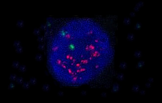
FISH/CISH
In situ hybridisation (fluorescent for FISH and chromogenic for CISH) uses probes to analyse genetic changes (amplifications, deletions, translocations) in tumours. These probes are used to classify tumours (particularly in the case of lymphomas and soft tissue tumours) and identify prognostic factors or therapeutic targets for certain tumours (in the case of breast cancers or lung adenocarcinomas).
Your contact person

PD DrDavide Soldini, Head Pathology
Head Pathology, Head molecular pathology
Specialist in Pathology and Molecular Pathology FMH

DrSimone Brandt
Deputy Lead Molecular Pathology
Specialist in Pathology and Molecular Pathology FMH


Our Molecular Pathology is an SIWF certified continuing education center.